Learn first: The excretory system and skin
The nervous system controls the work of all organs, links them together and connects the organism with its environment. Exam questions often cover the structure of the neuron, the reflex arc, the functions of the spinal cord and parts of the brain, and the effects of the sympathetic and parasympathetic systems. Be ready especially for "what is lost if this part is damaged" problems.
§1. Nervous tissue and the neuron
Nervous tissue consists of neurons and neuroglia cells, which nourish and protect them. The properties of a neuron are excitability and conductivity.
- Cell body — the part containing the nucleus.
- Dendrites — short branching processes; carry impulses towards the cell body.
- Axon — usually a single long process; carries impulses away from the cell body. Many axons have a myelin sheath, which speeds up conduction.
By function, neurons are of three kinds:
- Sensory (afferent) — carry impulses from receptors to the centre;
- Interneurons (relay neurons) — in the CNS, link sensory and motor neurons;
- Motor (efferent) — carry impulses from the centre to an effector (muscle or gland).
Clusters of neuron cell bodies form grey matter; myelinated fibres form white matter. A cluster of cell bodies outside the CNS is a ganglion.
A synapse is the junction between a neuron and another neuron or effector cell: presynaptic membrane, synaptic cleft, postsynaptic membrane. The impulse is transmitted by a chemical — a neurotransmitter (acetylcholine, noradrenaline and others).
At a synapse the impulse travels in one direction only: from the presynaptic to the postsynaptic membrane.
§2. Divisions of the nervous system
By structure:
- Central nervous system (CNS) — brain and spinal cord;
- Peripheral nervous system — nerves and ganglia.
By function:
- Somatic — controls skeletal muscles, voluntary movement;
- Autonomic — regulates internal organs, blood vessels and glands, usually without conscious control. It has two divisions.
The sympathetic division prepares the body for activity and stress: the heart beats faster and harder, the pupils dilate, the bronchi widen, gut movement and digestive secretion are reduced.
The parasympathetic division serves rest and recovery: the heart slows, the pupils constrict, digestion is stimulated.
Sympathetic centres lie in the thoracic and lumbar spinal cord; parasympathetic centres lie in the brainstem and the sacral spinal cord. The largest parasympathetic nerve is the vagus nerve (cranial nerve X).
§3. Reflexes and the spinal cord
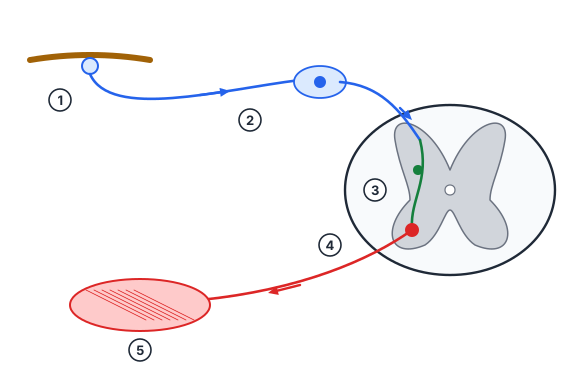
A reflex is the body’s response to a stimulus with the participation of the CNS. Its pathway is the reflex arc:
- Receptor — detects the stimulus.
- Sensory neuron — carries the impulse to the CNS.
- Interneuron — in the CNS (absent in some reflexes).
- Motor neuron — carries the impulse to the effector.
- Effector — a muscle or gland responds.
The simplest reflex arc has two neurons — for example, the knee-jerk reflex (no interneuron).
The spinal cord lies in the vertebral canal as a long white cord. In cross-section, butterfly-shaped grey matter is seen in the centre with white matter around it; the central canal runs through the middle. 31 pairs of spinal nerves leave the cord.
- Dorsal (sensory) roots — processes of sensory neurons; they bear the spinal ganglia.
- Ventral (motor) roots — axons of motor neurons in the ventral horns.
Functions of the spinal cord: reflex (knee-jerk, withdrawal reflexes, centres of urination and defecation) and conduction (carrying impulses to and from the brain).
Worked example
A patient feels a touch on the right leg but cannot move it. Which part of the spinal cord is damaged?
- Sensation is preserved → the sensory neuron and dorsal root are intact.
- There is no movement → impulses do not reach the muscle.
- Answer: the ventral (motor) root of that segment, or the motor neurons of the ventral horn, are damaged.
§4. The brain
The brain lies in the cranial cavity; 12 pairs of cranial nerves leave it. The brainstem consists of the medulla oblongata, the pons and the midbrain.
- Medulla oblongata — continuation of the spinal cord; vital centres of breathing, heart activity and blood vessels, protective reflexes (coughing, sneezing, vomiting), salivation and swallowing. Damage to it is life-threatening.
- Pons — links parts of the brain; contains nuclei of nerves involved in chewing and facial expression.
- Cerebellum — coordination of movement, balance, muscle tone. When it is damaged, gait becomes unsteady and movements lose precision.
- Midbrain — orienting reflexes to sudden light and sound, regulation of muscle tone.
- Diencephalon: the thalamus relays all sensory pathways (except smell) to the cortex; the hypothalamus controls body temperature, thirst, hunger and satiety, sleep and autonomic functions; it is linked to the pituitary gland and unites nervous and hormonal control.
- Cerebral hemispheres — covered by a cortex of grey matter; sulci and gyri increase its surface. The hemispheres are joined by the corpus callosum.
Lobes of the cortex: frontal — voluntary movement, thinking, the motor speech centre; parietal — skin and muscle sensation; temporal — hearing (the smell and taste areas lie nearby); occipital — vision.
Worked example
A drunk person moves clumsily and staggers. Which part of the brain is particularly affected by alcohol?
- Balance and coordination of movement are functions of the cerebellum.
- Answer: the cerebellum (though alcohol harms the whole CNS, especially the cortex).
§5. The nerve impulse, inhibition and care of the nervous system
A nerve impulse is an electrical signal (a wave of excitation) that travels along the membrane of a neuron. At rest the inside of the membrane is negative relative to the outside; when the neuron is excited, Na⁺ ions enter the cell and the charge at that point briefly reverses and then recovers. The change spreads to the next patch of membrane, so the impulse runs along the axon.
Impulse speed depends on the type of fibre:
- in thin unmyelinated fibres — about 0.5–2 m/s (many autonomic fibres to internal organs);
- in thick myelinated fibres — 100 m/s or more (motor fibres to skeletal muscles). Excitation arises only at gaps in the myelin sheath (nodes of Ranvier), so the impulse “jumps” from node to node.
Speed = distance / time. An impulse travels tens of times faster along a myelinated fibre than along an unmyelinated one.
Inhibition is an active process that weakens or stops excitation. Central inhibition was discovered by I. M. Sechenov (1862). Excitation and inhibition work together: when a leg bends, the centre of the flexor muscles is excited while the centre of the extensors is inhibited, so the movement is smooth.
Care of the nervous system: enough sleep (8–9 hours a day for teenagers), alternating mental and physical work, fresh air. Alcohol, nicotine and drugs disrupt transmission at synapses, damage neurons and cause dependence; the developing teenage brain is especially vulnerable.
Worked example
A motor fibre from the spinal cord to a toe muscle is 1 m long and conducts at 100 m/s. Compare this with the time over the same distance in an unmyelinated fibre (1 m/s).
- Myelinated fibre: t = 1 / 100 = 0.01 s (10 ms).
- Unmyelinated fibre: t = 1 / 1 = 1 s.
- Ratio: 1 / 0.01 = 100 times.
- Answer: 0.01 s in the myelinated fibre and 1 s in the unmyelinated one — myelin is essential for fast motor reflexes.
Check yourself
- What is the process that carries impulses away from the cell body called?
- How do the pupils and heart rate change when the sympathetic nerves are stimulated?
- Where are the breathing and heart centres located?
- In which lobe of the cortex is the visual area?
- How many neurons are in the knee-jerk reflex arc?
- What happens if a dorsal root of the spinal cord is cut?
- Who discovered central inhibition?
- An impulse travels at 50 m/s along a fibre 0.5 m long. How long does it take?
Common mistakes
- Writing that the sympathetic nerves slow the heart — they speed it up; the parasympathetic (vagus) nerve slows it.
- Placing the visual area in the temporal lobe — it is in the occipital lobe; the temporal lobe deals with hearing.
- Reversing the direction of conduction in dendrites and axons.
- Calling the dorsal root motor (dorsal is sensory, ventral is motor).
- Treating the cerebellum as the centre of thinking; thinking is a function of the cortex, especially the frontal lobe.
- Saying grey matter consists of myelinated fibres (myelinated fibres make up white matter).
Score: 0 / 8 correct (0 attempted)